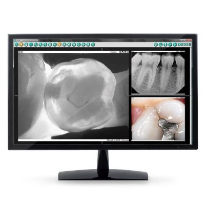
The software’s intuitive interface and its outstanding automation will let you breeze through X-ray procedures. Most common tasks are done in two clicks or less. Built-in X-ray sequences allow taking series with the fewest holder changes. With the introduction of DEXIS Eleven, your workflow is further simplified with drag-and-drop tooth numbering and the new history view which lets you sort a patient’s images by date for fast search and retrieval of a particualar image from the past.
When the sensor detects radiation, the image is automatically saved, dated, tooth numbered, correctly oriented and mounted, all without the need to return to the keyboard. Images are conveniently displayed in their anatomical location.
As a powerful, centralized imaging hub for all patient images, DEXIS manages all digital images, including intra and extraoral radiographs, as well as intra and extraoral photographs.
Stored images can instantly be organized, retrieved, printed and shared with patients, colleagues and insurance carriers. You can export and e-mail a full-mouth or bitewing series, including annotations, as individual files or as a single image.
DEXIS software is highly intuitive and easy to use. Most common procedures are done in 2 clicks or less. Its “1-Click Full-Mouth Series” makes it possible to reduce a 25-minute FMX procedure to just 5 minutes, start to finish.*
When the sensor detects radiation, the image is automatically saved, dated, tooth numbered, correctly oriented and mounted, all without the need to return to the keyboard. Built-in X-ray sequences allow taking series with the fewest holder changes.*
Images are available instantly after exposure, eliminating the wait and effort spent developing and mounting X-rays. If an image needs to be retaken, it can be done immediately. Large onscreen X-rays make patient communication more effective too.
The magnification feature, along with a comprehensive set of image enhancement tools, provide support for clinical diagnosis, including the identification of apical and carious lesions, open margins, bone loss and furcation involvement.
As a powerful imaging hub, DEXIS manages all digital images, including intra- and extra-oral radiographs, as well as intra and extraoral photos. Stored images can instantly be organized, retrieved, printed and shared with patients, colleagues and insurance carriers.
This innovative method of image processing optimizes the signal path between image acquisition and on-screen display resulting in clear, highly detailed radiographs.
The ability to see subtleties in X-rays is crucial to diagnosis, collaboration and patient communication. DEXIS hardware and software work in harmony to deliver the highest quality, most consistent images at the widest range of exposure settings.1
ClearVu™, DEXIS’ acclaimed image enhancement tool, uses advanced algorithms to further define the radiograph, resulting in clinically meaningful images that are sharp, detailed and rich in contrast.
Export a full-mouth or bitewing series, including annotations, as individual files or as a single image for convenient sharing and collaboration via e-mail. JPEG and TIFF formats are supported as well as DEX format.
DEXIS accommodates the specific workflows of general dentistry and specialties including endodontics, periodontics, prosthodontics and oral surgery.
| CPU | Intel® Core™ i3 Processor or higher |
| Motherboard | Intel chipsets |
Operating System | |
| Workstations | Windows® 7 (32-bit) Professional, Ultimate, Enterprise – SP1 Windows 7 (64-bit)* Professional – SP1 Windows 8.1 (32/64-bit) Windows 10 (32/64-bit)
*IMPORTANT Note about Windows 64-bit: Not all hardware is 64-bit compatible, it is crucial to evaluate all digital hardware in your practice for 64-bit compatibility prior to purchasing or upgrading your PC(s) to 64-bit. |
| Servers | Windows Server 2008 – SP2 Windows Server 2008 R2 – SP1 Windows Server 2012 R2 Note: Dedicated file servers above are recommended in networks with more than 5 imaging workstations.
|
| Microsoft SQL | Microsoft® SQL Server 2005 (Express version supplied with software) Microsoft SQL Server 2008 Note: Express, Standard and Enterprise versions supported. |
System Memory | |
| Workstations | 4 GB or higher |
| Servers | 8 GB or higher |
Hard Disk Drive | |
| Workstations | 160 GB or larger |
| Servers | 500 GB or larger |
Graphics Card |
Capable of displaying a minimum of 1024 x 768 pixels in 24-bit color or higher |
| Monitor | 21″ High resolution widescreen LCD with a contrast ratio of 10,000:1 or better Note: LCD monitors should be used in native resolution, and must display all shades of gray accurately. |
| USB | USB 2.0, USB 3.0 |
| Network Card | 100/1000 baseT network cards Note: Wireless networks are not recommended. |
| DVD Drive | 16x or higher. Required to install the DEXIS software. |
| Mac Compatibility | Mac® OS X® Snow Leopard 10.6 or higher running Parallels® version 5 or higher and an approved Windows operating system (see “Operating System” above). |
| Supported Hardware | Visit www.dexis.com/hardware for a detailed list of currently tested and supported hardware. |
DEXIS Imaging Suite next generation software architecture brings exciting new features including a cosmetic imaging module, an enhanced planning module, added video capabilities in DEXimage and integration with select 3D products.

DEXvoice is an exciting NEW add-on module to the DEXIS imaging software that allows customers to use any Amazon Alexa enabled platform to integrate voice activation to their dental workflow.
Enable dental practices to improve productivity during image acquisition and patient presentation, keeping their attention on patients and not the computer.

Amazon, Alexa and all related logos are trademarks of Amazon.com, Inc. or its affiliates.

View, capture and manage intraoral and extraoral camera images from a digital photo camera, intraoral HD video camera or microscope. Live Video offers full video recording and playback capabilities as well as the ability to capture still images from recorded video.

This module greatly simplifies implant planning and selection with an extensive library of implant models. It provides a visual aid in determining placement near anatomic structures and long-term assessment of bone integration.

Connect DEXIS, the 3D i-CAT scanner, and KaVo ORTHOPANTOMOGRAPH™ OP 3D & ORTHOPANTOMOGRAPH OP 3D PRO with the all-new DEXIS i-CAT & KaVo Link. Seamlessly manage patient data and 3D images from 3D scanners directly from within the DEXIS application.

Customizable chairside report writer lets you ‘drag and drop’ intra and extraoral X-rays and photo- graphic images into a document. Use clinical templates or create your own letters and marketing materials in minutes.

Quickly and easily configure and attach X-rays, pans, photos, perio charts, EOBs, treatment cards, etc., in support of insurance claims and send them electronically for immediate review by insurance carriers.

Multi-user, multi-computer site license for operating the DEXIS system(s) and software modules on an unlimited number of locally networked workstations.

非常抱歉,您只有购买软件后才能查看完整软件教程!
| 版本号 | 软件大小 | 下载地址 |
|---|
| 版本号 | 软件大小 | 下载地址 |
|---|

