Temporomandibular joint ankylosis, or the fusion of the jawbone, is most often the result of an injury or infection, and it prevents the patient from opening their mouth properly. It can only be treated with surgery, but due to the complex nature of the operation which presents a lot of risks for the patient, surgeons are often too careful to really perform an effective surgery. This means that the problem isn’t solved properly and is in danger of recurring.
The development of a new type of piezoelectric saw has presented surgeons with a novel way of safely performing the surgery, as it claims to do no damage to soft tissues. It also prevents dural tears, the perforation of the external auditory canal and bleeding from the maxillary artery and inferior alveolar vascular bundle.

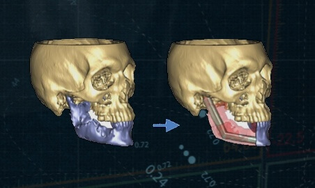
3D model for temporomandibular joint ankylosis surgery
Dr. Bhaskar Agarwal and his team used Materialise PROPLAN CMF software to reconstruct a computed tomography for one of their patients. The patient in question was a 20-year-old female, who had been unable to open her mouth for the last ten years – although she had previously undergone surgery to treat the disease, the condition recurred. The most common causes of temporomandibular joint ankylosis are infection, trauma and birth defects and in the case of the patient, she had experienced trauma to the mandible, which led to a mandibular fracture which wasn’t treated properly. Since her condition was recurring, it was necessary for her to undergo aggressive bone removal in order to create a large enough gap in her jaw.
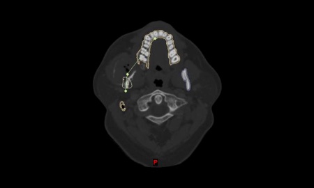
Using the model on the computer, they decided where to position the superior and inferior cuts on the patient’s skull. They then printed out the model of the skull in 3D using Stereolithography, and featured projecting osteotomy planes on the ankylosed bony mass. As well as planning the cuts, the team used the virtual model to create sterilized surgical guides. This would enable the surgeon to make precise cuts during the operation. The guides used during the procedure were traditionally manufactured, using a biocompatible self-curing polymethyl methacrylate resin. However, they can alternatively be made just as effectively with 3D Printing technology.

3D-printed model with surgical guides in place
During the temporomandibular joint ankylosis surgery, the team implemented these surgical guides and the piezoelectric saw. Using micro movements, the saw ensured that the bone debris did not build up and prevent the binding of the instrument during the osteotomy. Then they removed the ankylotic mass using the surgical cutting guides. The maxillary artery and inferior alveolar vascular bundle were close to the bony mass. However, the precision of the cutting guides allowed the surgeons to safely remove it without damaging any of the tissues or arteries, making the operation a success.
Dr. Bhaskar Agarwal
Dr. Bhaskar Agarwal is a Resident Doctor in the Department of Oral and Maxillofacial Surgery at the All India Institute of Medical Sciences in New Delhi. His area of interests are cranio-maxillofacial deformities, traumatic injuries to the craniofacial skeleton and pathologic diseases of the jaw. He is a firm believer in virtual surgical planning for the cranio-maxillofacial region, and believes that technological advancements in the field of surgery have the potential to make cranio-maxillofacial surgery safer and more predictable. Read publications of Dr. Bhaskar Agarwal

非常抱歉,您只有购买软件后才能查看完整软件教程!
| 版本号 | 软件大小 | 下载地址 |
|---|
| 版本号 | 软件大小 | 下载地址 |
|---|


